Home » Without Label » Abdominal Anatomy - Arteries of the abdomen at University of Oregon - StudyBlue - This might sound like a strange question, right?
Abdominal Anatomy - Arteries of the abdomen at University of Oregon - StudyBlue - This might sound like a strange question, right?
Abdominal Anatomy - Arteries of the abdomen at University of Oregon - StudyBlue - This might sound like a strange question, right?. The rectus abdominis connects to the xiphoid process, a bony landmark at the bottom of the sternum. You can't have a strong, muscular physique without a healthy, stable core. The major organs of the abdomen include the small intestine, large intestine, and stomach. Learn the anatomy and function of your abdominals to achieve your dream physique. The anterolateral abdominal wall formed of 4 layer skin, fascia, muscles, and peritoneum.
Divided into 9 regions by two vertical and two horizontal imaginary planes. Abdomen, in human anatomy, the body cavity lying between the chest or thorax above and the pelvis below and from the spine in the back to the wall of abdominal muscles in the front. The area occupied by the abdomen is called the abdominal cavity. This might sound like a strange question, right? Assoc prof craig hacking and dr pradeep a wijayagoonawardana et al.
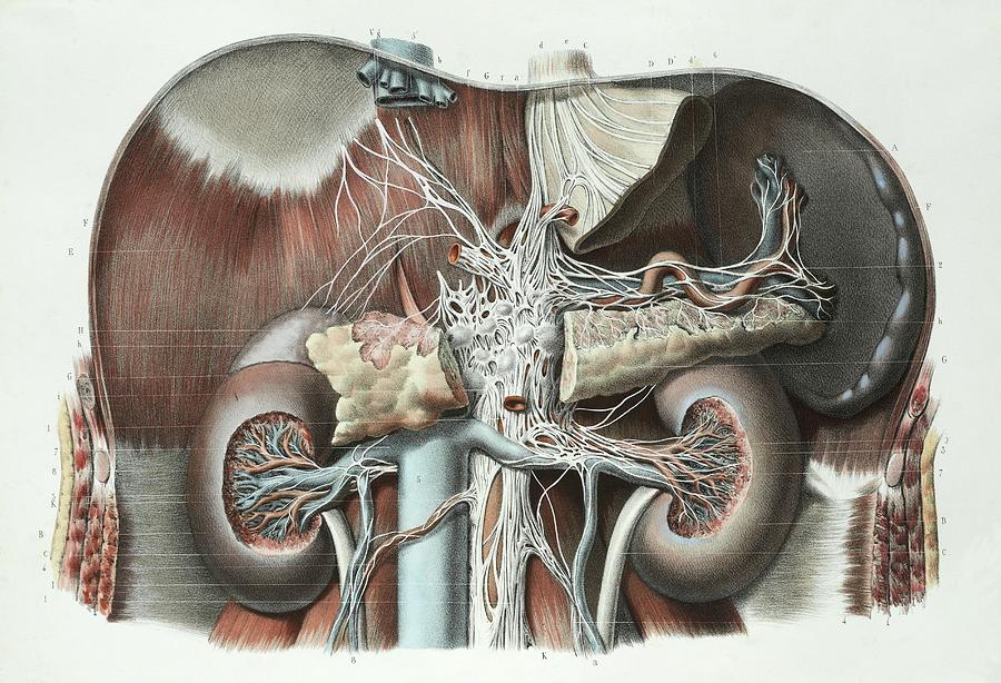
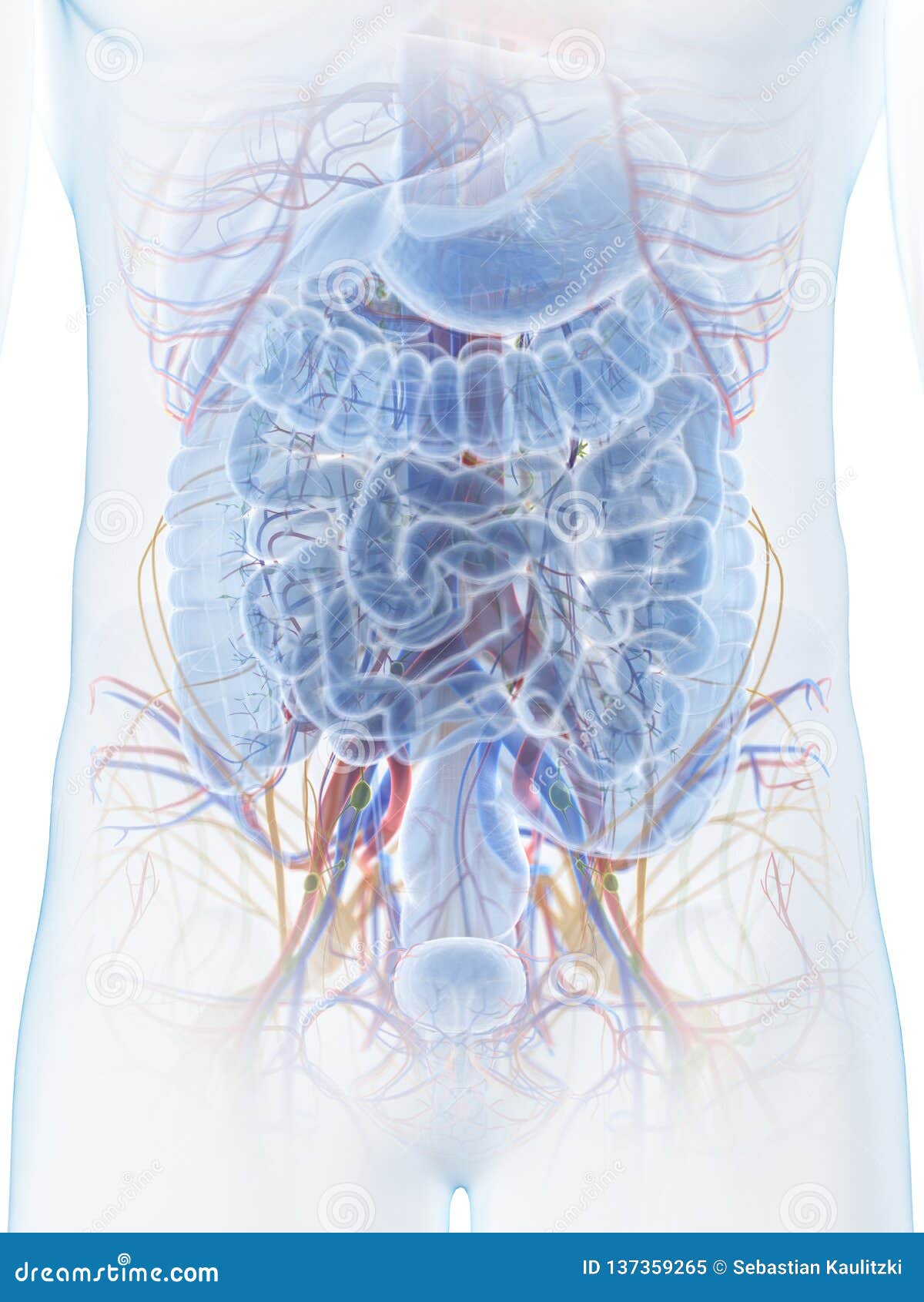
Abdominal Anatomy Photograph by Science Photo Library from images.fineartamerica.com The image also shows the pelvis, uterus, and urinary. It is bounded superiorly by the xiphoid process and costal margins, posteriorly by the vertebral column and inferiorly by the pelvic bones and inguinal ligament. The liver, stomach, and abdominal contents are clearly identified and labeled, including the cecum, ascending colon, transverse colon, descending colon, and small intestine. The aorta is the largest blood vessel in the body. The area occupied by the abdomen is called the abdominal cavity. Abdominal surface anatomy can be described when viewed from in front of the abdomen in 2 ways: Let's learn more about the anatomy of the abdomen. These organs are held together loosely by connecting tissues.
The anterolateral abdominal wall consists of four main layers (external to internal):
You can't have a strong, muscular physique without a healthy, stable core. Inferiorly the abdomen is open to the pelvis, communicating through the superior pelvic aperture (pelvic inlet). Skin, superficial fascia, muscles and associated fascia, and parietal peritoneum. It is a body space situated between the thorax and the pelvis. The anterolateral abdominal wall consists of four main layers (external to internal): These two apertures, together with abdominal walls, bound the abdominal cavity. Together, these three turn nutrients into usable energy, as well as help dispose of solid waste. Let's learn more about the anatomy of the abdomen. The rectus abdominis connects to the xiphoid process, a bony landmark at the bottom of the sternum. It is an artery, meaning that it carries blood away from the heart. The abdomen (colloquially called the belly, tummy, midriff or stomach) is the part of the body between the thorax (chest) and pelvis, in humans and in other vertebrates. It is the long, flat muscle that extends vertically between the pubis and the fifth, sixth, and seventh ribs. The abdomen is the front part of the abdominal segment of the trunk.
We'll identify as many organs as we can, see how they fit into the. The regions occupied by stomach are epigastric, umbilical and hypochondriac regions. It is a body space situated between the thorax and the pelvis. If you plan to enter a healthcare profession such as nursing, this is something you'll use on the job when performing abdominal assessments (and while documenting). Use the mouse scroll wheel to move the images up and down alternatively use the tiny arrows (>>) on both side of the image to move the images.>>) on both side of the image to move the images.
The abdominal anatomy stock illustration. Illustration of ... from thumbs.dreamstime.com Muscles of the anterior abdominal wall consists of two vertical muscles located on the midline and bisected by linea alba; Stomach is a muscular bag forming the most distensible part of the human digestive system. Abdomen anatomy the abdomen is comprised primarily of the digestive tract and other accessory organs which assist in digestion, the urinary system, spleen, and the abdominal muscles (shown below). The region occupied by the abdomen is called the abdominal cavity, and is enclosed by the abdominal muscles at front and to the sides, and by part of the vertebral column at the back. The abdomen (colloquially called the belly, tummy, midriff or stomach) is the part of the body between the thorax (chest) and pelvis, in humans and in other vertebrates. Connective tissue called the mesentery holds the abdominal organs together. Abdominal surface anatomy can be described when viewed from in front of the abdomen in 2 ways: Divided into 9 regions by two vertical and two horizontal imaginary planes.
It is an artery, meaning that it carries blood away from the heart.
These organs are held together loosely by connecting tissues. The diaphragm is its upper boundary. Together, these three turn nutrients into usable energy, as well as help dispose of solid waste. Divided into 4 quadrants by single vertical and horizontal imaginary planes. It is a body space situated between the thorax and the pelvis. The abdomen contains all the digestive organs, including the stomach, small and large intestines, pancreas, liver, and gallbladder. It is the long, flat muscle that extends vertically between the pubis and the fifth, sixth, and seventh ribs. The abdomen is the body region found between the thorax and the pelvis. If you plan to enter a healthcare profession such as nursing, this is something you'll use on the job when performing abdominal assessments (and while documenting). The rectus abdominis connects to the xiphoid process, a bony landmark at the bottom of the sternum. Divided into 9 regions by two vertical and two horizontal imaginary planes. The liver, stomach, and abdominal contents are clearly identified and labeled, including the cecum, ascending colon, transverse colon, descending colon, and small intestine. The abdominal cavity is the part of the body that houses the stomach, liver, pancreas, kidneys, gallbladder, spleen, and the large and small intestines.the diaphragm marks the top of the abdomen and the horizontal line at the level of the top of the pelvis marks the bottom.
The diaphragm is its upper boundary. We're going to take apart a plastic anatomy model and see what we can find in the abdomen. It is a body space situated between the thorax and the pelvis. Muscles of the anterior abdominal wall consists of two vertical muscles located on the midline and bisected by linea alba; Rectus abdominis and pyramidalis and three flat muscles on the anterolateral side arranged from superficial to deep;
The abdominal anatomy stock illustration. Illustration of ... from thumbs.dreamstime.com The aorta is the largest blood vessel in the body. Its superior aperture faces towards the thorax, enclosed by the diaphragm. When you think of abs, what muscle do you typically think of? Divided into 9 regions by two vertical and two horizontal imaginary planes. The abdomen is the part of the body that contains all of the structures between the thorax (chest) and the pelvis, and is separated from the thorax via the diaphragm. The anterolateral abdominal wall consists of four main layers (external to internal): The regions occupied by stomach are epigastric, umbilical and hypochondriac regions. The abdomen contains all the digestive organs, including the stomach, small and large intestines, pancreas, liver, and gallbladder.
The aorta is the largest blood vessel in the body.
Abdomen anatomy the abdomen is comprised primarily of the digestive tract and other accessory organs which assist in digestion, the urinary system, spleen, and the abdominal muscles (shown below). Muscles of the anterior abdominal wall consists of two vertical muscles located on the midline and bisected by linea alba; In anatomy and physiology, you'll learn how to divide the abdomen into nine different regions and four different quadrants. The abdomen is the body region found between the thorax and the pelvis. Use the mouse scroll wheel to move the images up and down alternatively use the tiny arrows (>>) on both side of the image to move the images.>>) on both side of the image to move the images. This mri abdomen axial cross sectional anatomy tool is absolutely free to use. The area occupied by the abdomen is called the abdominal cavity. Learn the anatomy and function of your abdominals to achieve your dream physique. The major organs of the abdomen include the small intestine, large intestine, and stomach. These organs are held together loosely by connecting tissues. You go to the gym to train your abs. This might sound like a strange question, right? Let's learn more about the anatomy of the abdomen.